Models of Health

We live in three dimensions, so should healthcare data.

Moving Beyond Flatland

  • 3D medical imaging provides context and diagnostic value by giving clinicians more than a flat stack of images.
  • When combined with additional detail, which itself can be pulled from the models; it is possible to create powerful visualizations, improve diagnostic accuracy or more effectively educate patients.
CT: Head Slice 1
CT and MRI scanners acquire data as two dimensional slices.
CT: Head Slice 2
Slices are stacked together into volumes which are then analyzed to find patterns.
Anatomic Models (3D): Skull and Neck
The models can be combined with the segmented data to provide physicians and patients with new ways of understanding their health and providing care.

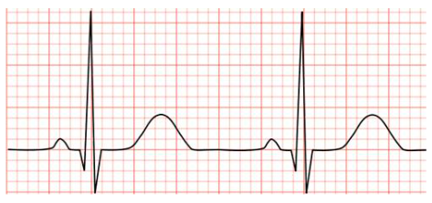
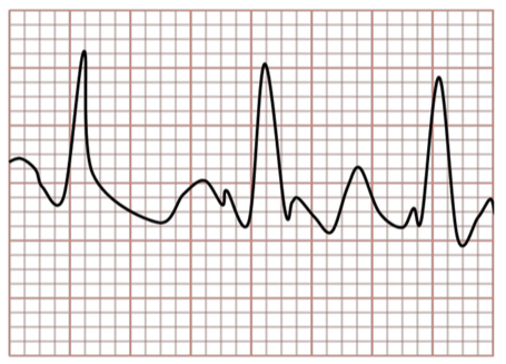
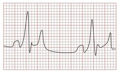
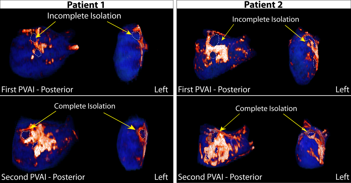
Assessing Atrial Fibrillation Treatment Efficacy

Machine vision techniques can provide tremendous insight about how to help cardiac arrhythmia patients.

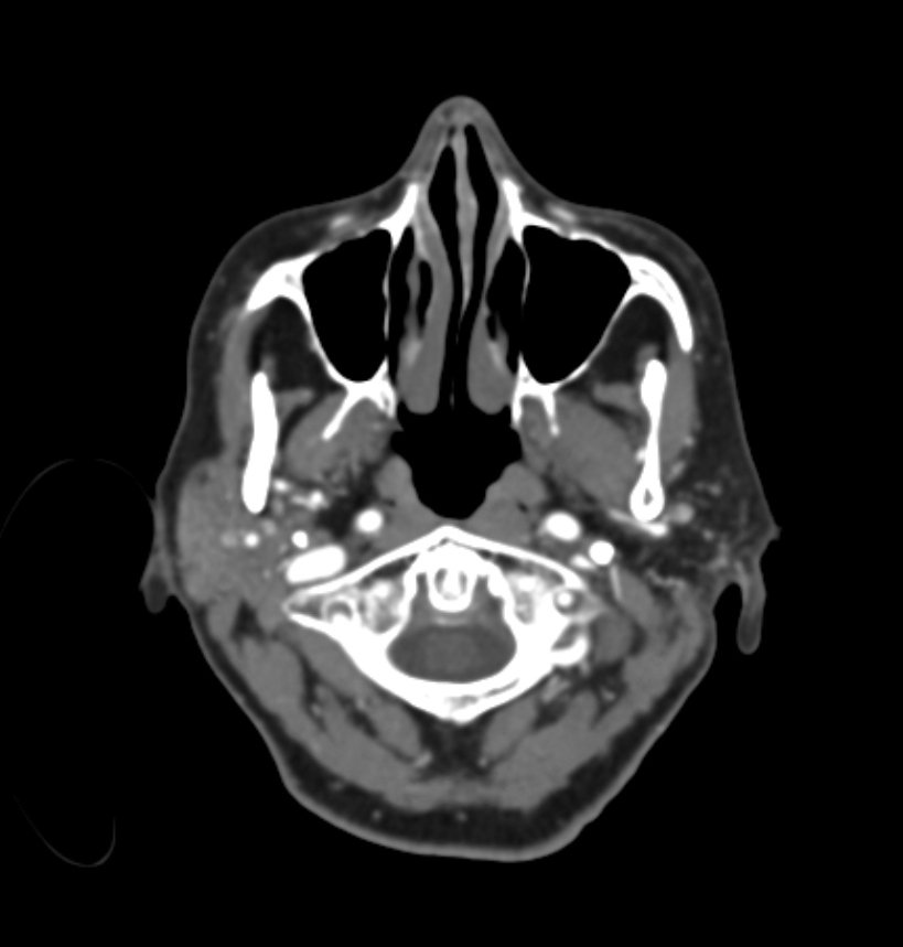
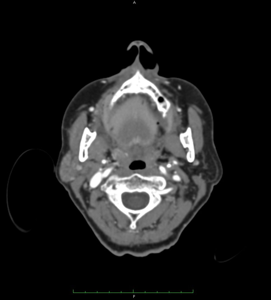
Atrial fibrillation (AF) is the most common arrhythmia in the world and impacts tens of millions of people. Patients suffering from AF live a diminished quality of life and are at risk for other serious conditions such as stroke.

EKG Trace: Healthy
A healthy heart has a coordinated and regular heart beat. Arrhythmias occur when parts of the heart lose their coordination due to errant electrical signals.
EKG Trace: Atrial Fibrillation
Atrial brillation is caused by uncoordinated electrical activity in the left atrium. It leads to an irregular heart beat which prevents the heart from effectively pumping blood.
EKG Trace: EPR
Atrial fibrillation is just one of many types of arrhythmias, but it can lead to others (such as elongated PR, shown above) and puts patients at risk for other health risks including heart attack and stroke.
Atrial Fibrillation: Assessing Patient Response to Ablation
To effectively stop atrial fibrillation, it is necessary to isolate electrical signals in the left atrium. Incompletely isolating the signals results in failed treatment.

Assess treatment early and effectively.

Atrial fibrillation is treated by finding unhealthy tissue and ablating (killing it)

  • Ablation treatment is a fine art form. Frequently, tissue is only partially ablated and will continue to contribute to uncoordinated electrical activity.
  • Knowing if tissue has been completely treated can be difficult to to assess without an invasive procedure.
  • Non-invasive (medical imaging) therapies combine with machine vision can help to quantify how effective a treatment has been and whether additional intervention is necessary.
Atrial Fibrillation: Building 3D Models from 2D MRI (Shows Ablation Scarring)
High contrast medical imagery combined with machine vision and segmentation techniques greatly facilitate the assessment of treatment efficacy. It can lead to the building of patient specific models to guide further intervention.
Atrial Fibrillation: Building 3D Models from 2D MRI (Segmentation Masks)

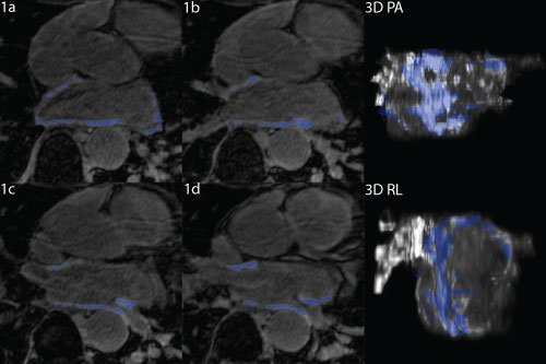
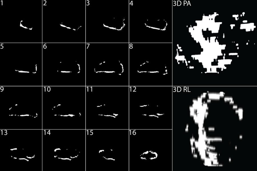
Machine learning allows the building of patient specific models.

It can be difficult to apply new therapies because of how labor intensive it can be to create models outside of the research lab. Machine learning can be leveraged to segment images rapidly and efficiently without losing accuracy.

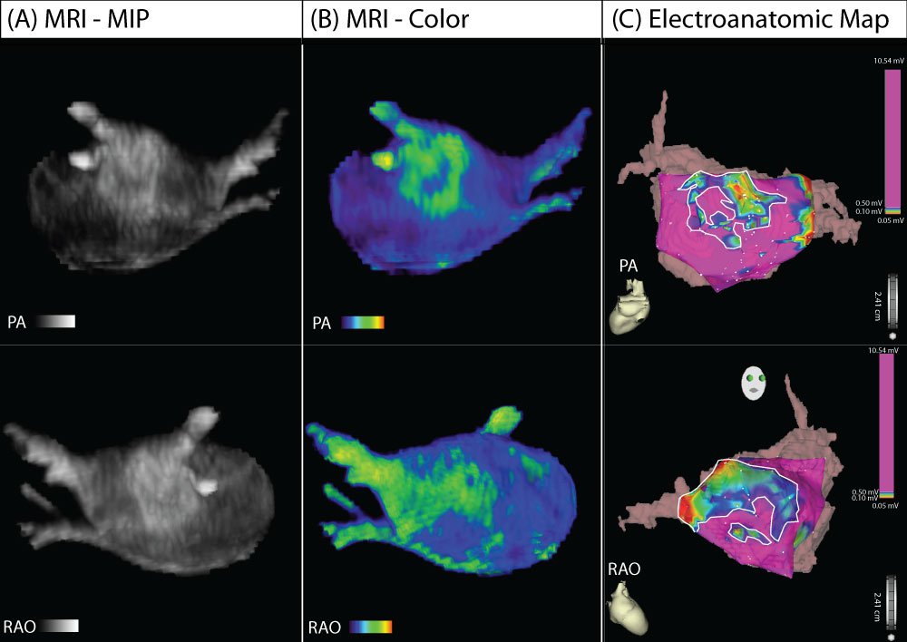
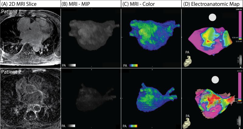
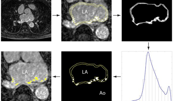
Quantifying the Severity of Atrial Fibrillation

Atrial brillation actively changes the structure of the heart, entrenching the arrhythmia and making it more difficult to treat.

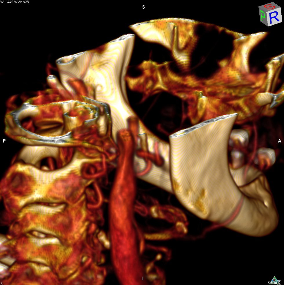
Structural changes in the heart correlate with electrical changes.

  • These structural changes aren't always reflected in the way the disease manifests; but strongly predict response to therapy.
  • They are really hard to see without an invasive study, though. Medical imaging alongside computer vision, segmentation, and machine learning assessment can give insight to how far along the disease really is. This information then informs treatment options leading to better outcomes.
Atrial Fibrillation: Structural as Compared to Electrical Change (Patient 1)
MRI studies (A) combined with machine vision techniques (B, C) can create patient specificc models of disease that are predictive of electrical abnormalities (D).
Atrial Fibrillation: Structural Versus Electrical Change (Patient 2)

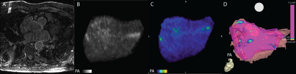
Pictures worth thousands of words.

MRI combined with image processing pipelines provide a powerful tool to inform doctors how to treat arrhythmias. Visualizations can help providers plan intervention and then to communicate findings and care plans to the patient.

Atrial Fibrillation: Structural Versus Electrical Change (Patients 3 and 4)

Creating positive feedback loops.

  • The resulting models provide key insights into how a patient will respond to treatment. Patients with more observed structural change and higher degree of scarring require much more aggressive therapy then those with less change.
  • Over time, as more data is acquired, it is possible to even more effectively target therapy and predict outcome.

Learn More

Comments

Loading
Unable to retrieve data due to an error
Retry
No results found
Back to All Comments